b value mri
Applications and Challenges in Oncology. Depending on the organ being imaged b-values typically range from 50-1000smm 2.

Diffusion Weighted Imaging Radiology Reference Article Radiopaedia Org
DBS Deep brain stimulation.
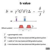
. Consequently when all other imaging parameters are held constant b. B γ² G² δ² Δδ3 Therefore a larger b value is achieved by increasing the gradient amplitude and duration and by widening the interval between paired gradient pulses. In all 67 healthy participants were divided into three age groups group A 15-30 years.
Multiple b values were used 50 400 800 and 1000smm2 at least 2 b values. To sense slow moving water molecules and smaller diffusion. Units are micro-Tesla µT.
The term b -value characterizes the diffusion gradient pulses amplitude shape and timing and expresses the amount of diffusion weighting 1. Also may be called the turbo factor. Most prior diffusion-weighted imaging studies of human brain infarction have been performed with b values of 1000 or less 2126 although one reported a b value of 1463 13.
The whole brain MRI examinations were performed on a 30-T MRI system Discovery MR750 GE Healthcare Milwaukee WI USA with a 40-mTm maximum gradient capability and an eight-channel head coil GE Medical Systems. Conventional MRI sequence and five consecutive multiple b-value DWI sequences of brain were performed during one. Group B 31-50 years.
Refocusing Pulse The spin-echo RF pulse used to refocus the MR signal. High-b value diffusion-weighted MRI DWI is different from morphologically oriented imaging techniques in that it can sensitively depict disease-associated changes of random translational molecular motion known as diffusion or brownian water motion. DWI and ADC value of.
ETL Echo train length. The b factor summarizes the influence of the gradients on the diffusion weighted images. DWI is done to determine the rate of molecular diffusion in different areas of the body.
The best b-value combination was 0 and 800 Az0935. There is no consensus on the optimal b-value. In general in healthy tissue molecules of water and other chemicals are not stationary but moving about.
Lesions were analyzed for benignitymalignity using apparent diffusion coefficient ADC values with 10 b-value combinations and by measuring the lesionnormal parenchyma ADC ratio. Group C 51 years and underwent DWI scanning twice with 15 b-factors from 0. Thus image sequences from low to high b-values must be read side by side in order to establish the precise anatomic location of the suspected tumor.
Diffusion-Weighted MRI in the Body. DT-MRI data were acquired along 19 unique gradient axes with a b- value of 1000 smm 2 and an additional image at b 0 smm 2 with the following parameters. B1rms The root-mean-square value of the MRI effective component of the B1 field.
Mean ADC values were approximately 13 1-15 higher in total when b0 and b50 smm 2 were included in multiple b-value combinations. To investigate the reproducibility of diffusion-weighted imaging DWI with ultrahigh b-values and analyze the age-related differences in normal prostates. The reason is readily apparent from the images below.
MATERIALS AND METHODS Patients. SAR Specific absorption rate. With b 0 bright signals are noted in multiple veins due to the high T2 of blood coupled with sluggish flow.
Certa in illnesses show restrictions of diffusion for example demyel in ization and cytotoxic edema. The study included 60 patients 34 male and 26 female with solid head and neck masses 1cm who referred to radiodiagnosis department for MRI evaluation. Modern scanners usually allow a range of 0-4000smm 2.
Routine abdominal MRI and DWI were performed using seven b-values 0 50 200 400 600 800 1000 smm 2. Diffusion magnetic resonance imaging dMRI is a unique technique to probe the microstructure of the normal and diseased tissue by quantifying the displacements of water molecules 1. The aim of our study was therefore to determine with the aid of histopathological findings for radical prostatectomy as a reference which bvalue b 1000 smm 2 or b 2000 smm 2 used for 3T MRI is more suitable for visual assessment for the detection of prostate cancer on native DWI.
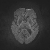
Brain DW images obtained at b 3000 appear significantly different from those obtained at b 1000 reflecting expected loss of signal from all areas of brain in proportion to their ADC values. A b value of 8001000 smm 2 would provide an excellent spatial resolution and an adequate signalnoise ratio for lesion evaluation. The degree of diffusion weight in g correlates with the strength of the diffusion gradients characterized b y the b - value which is a function of the gradient related parameters.
The higher the value b the stronger the diffusion weighting. Strength duration and the period b etween diffusion gradients. The b-value is a factor of diffusion weighted sequences.
The b value is used in MRI in the context of Diffusion Weighted Imaging DWI. These studies used different b values varying from 0 to 1000 smm 2 and found a significant difference of the ADC value between malignant and benign lesions with a sensitivity ranging from 81 to 93 and specificity from 80 to 88 for an ADC cutoff of 1113 10 3 mm 2 s. Using lower b-values such as 0 and 50 together with higher b-values 600 smm 2 was beneficial Az0928 and 0927.
The purpose of this study was to determine the role of high-b-value b 2500 or 3000 diffusion-weighted imaging for lesion detection in acute and chronic brain infarction. The use of b values more than 1000 smm 2 would offer better contrast but was more liable to suffer susceptibility artifact. Pulse repetition timeecho time 1500091 ms three averages number of excitations field of view 202 cm matrix size 128 128 giving a resolution of 158 158 21 mm 3.
A baseline b-value of 50 smm² is often used in liver diffusion-weighted imaging instead of b 0.

Diffusion Weighted Imaging In Acute Ischemic Stroke Radiology Reference Article Radiopaedia Org

Hyperpolarised 13c Mri Identifies The Emergence Of A Glycolytic Cell Population Within Intermediate Risk Human Prostate Cancer Nature Communications

Signal Intensity Of Dwi And Adc In Diffusion Restriction Increased Download Scientific Diagram

Diffusion Weighted Imaging Radiology Reference Article Radiopaedia Org

Diffusion Tensor Imaging And Fiber Tractography Radiology Reference Article Radiopaedia Org
Tensor Valued Diffusion Encoding For Diffusional Variance Decomposition Divide Technical Feasibility In Clinical Mri Systems Plos One

Cerebral Micro Structural Changes In Covid 19 Patients An Mri Based 3 Month Follow Up Study Eclinicalmedicine
Tensor Valued Diffusion Encoding For Diffusional Variance Decomposition Divide Technical Feasibility In Clinical Mri Systems Plos One

Diffusion Weighted Imaging Radiology Reference Article Radiopaedia Org

Diffusion Weighted Imaging Radiology Reference Article Radiopaedia Org

Principles Of Diffusion Tensor Imaging And Its Applications To Basic Neuroscience Research Neuron

Apparent Diffusion Coefficient Radiology Reference Article Radiopaedia Org

Apparent Diffusion Coefficient Radiology Reference Article Radiopaedia Org

Principles Of Diffusion Tensor Imaging And Its Applications To Basic Neuroscience Research Neuron
0 Response to "b value mri"
Post a Comment